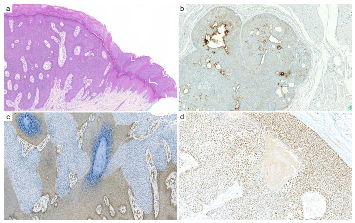
Figure 6.
Eccrine poroma: (a) cutaneous eccrine poroma is composed of a population of poroid cells, with a sharp demarcation with adjacent epidermis, inconspicuous ducts, and numerous dilated vessels in the papillary dermis (×25); (b) CEA immunohistochemistry highlights the ductal differentiation (×200); (c) YAP1 (c-terminal) immunohistochemistry shows a clonal loss of expression in poroid cells compared to the adjacent epidermis, which suggest a YAP1-fusion (×200); (d) NUT immunohistochemistry demonstrates diffuse nuclear staining when a NUTM1 gene fusion is involved, which is more frequent in the poroid hidradenoma variant (×200).

