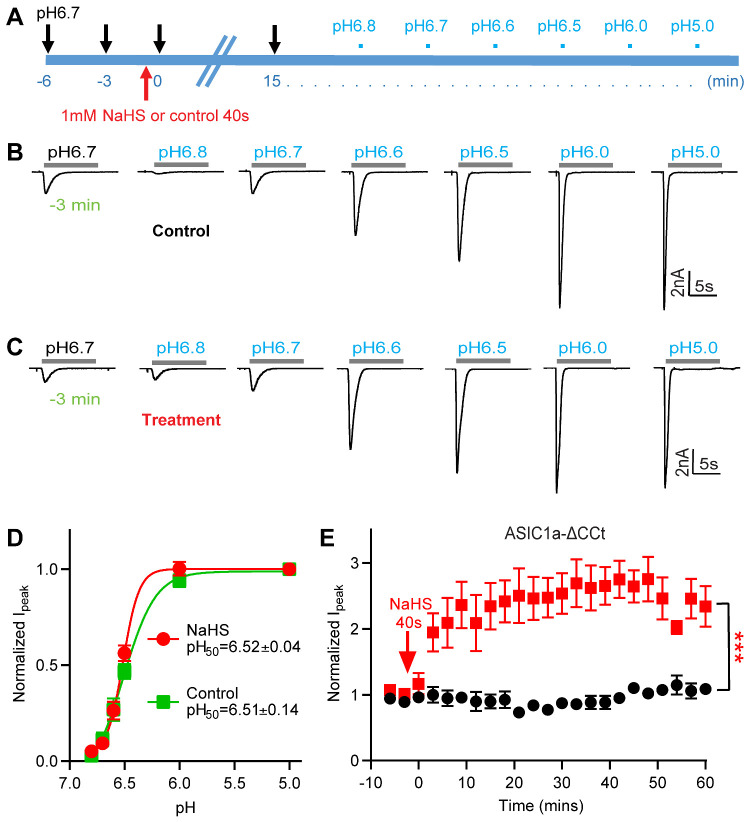
Figure 3.
NaHS Potentiation of ASIC1a Currents Is Not Due to a Change in pH Dependence. (A) Schematic representation of the protocol used to test whether the exposure to 1 mM NaHS induces a shift in the pH dependence of ASIC1a expressed in CHO cells. (B and C) Representative ASIC1a current traces for the construction of a pH–response curve. Fifteen minutes before starting the recording of the pH-response curve, the cell was exposed during 40 s to a control solution (B, “control”) or to a solution containing 1mM NaHS (C, “treatment”). (D) ASIC1a peak current amplitudes, normalized to the peak amplitude induced by pH 5.0, for cells exposed to 1 mM NaHS (treatment, red) or not (control, green), n = 5–9. The solid lines represent a fit to the Hill equation. The pH50 values were not different between the two conditions (unpaired Student’s t-test). (E) Time course of the pH 6.7-induced current of CHO cells expressing a mutant ASIC1a in which the intracellular C-terminal Cys residues were mutated or deleted (ASIC1a-C466A-C471A-C497A-C528stop, ASIC1a-ΔCCt), measured without (control, black symbols) or with a 40-s exposure to 1 mM NaHS at time point 0, as indicated (treatment, red symbols), n = 4–6. ***P < 0.001, comparison between treatment and control over the period 0–60 min by one-way ANOVA test and Dunnett’s post hoc test. Current amplitudes were normalized to the pH 6.7-induced current amplitude measured before NaHS exposure (at −3 and −6 min).

