Figure 5.
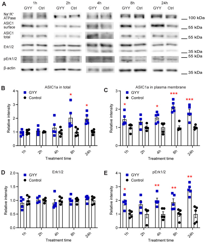
H2S Donors Regulate the Expression of ASIC1a and the Activation of the Erk1/2 Signaling Pathway. The biochemical experiments were carried out in cultured mouse cortical neurons. Total and plasma membrane proteins were isolated, separated on SDS-PAGE, and specific proteins were visualized as described in the “Materials and Methods” section. (A) Representative Western blots of total and plasma membrane ASIC1a, and Erk1/2, p-Erk1/2, Na+/K+ATPase, and β-actin as indicated, after incubation with 10 µM GYY4137 (GYY) or without (Ctrl) for the indicated time. β-actin was used as a control for the total protein, and Na+/K+ ATPase α1 as a control for plasma membrane proteins. The β-actin and Na+/K+ ATPase bands shown in (A) were from the same sample, but not in all cases from the same lane on the gel, as the bands shown above or below. (B–E) Cells were exposed to 10 µM GYY4137 (GYY, blue symbols) or to control medium (control, black symbols) for the indicated time. The measured intensities were normalized to the average intensity of the corresponding control. (B) Total expression of ASIC1a, n = 4–5. (C) Plasma membrane expression of ASIC1a, n = 4–5. (D) Expression of Erk1/2, n = 4–5. (E) Expression of p-Erk1/2, n = 4–5. *P < 0.05; **P < 0.01; comparison of each treatment condition with the corresponding control condition by multiple Mann–Whitney tests.

