
FIGURE 1.
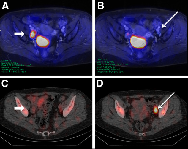
A 58-y-old woman with newly diagnosed T2b G3 cervical carcinoma. PET/MRI (top row) detected enlarged LN in right hemipelvis (A, arrow) with intense 18F-FDG uptake. In contrast, 99mTc-nanocolloid SPECT/CT showed clearly definable SLN in left hemipelvis (D, arrow) with benign characteristics on PET/MRI (B). After surgery, 1 LN in right hemipelvis (A and C) was confirmed as LNM. SLN on left (B and D) had benign histology.