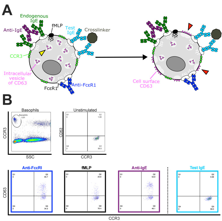
Figure 4.
Example of the basophil activation test (BAT) to evaluate the propensity of an anti-FRα IgE antibody (MOv18 IgE) to trigger human basophil activation. (A) Schematic of basophil containing intracellular vesicles of CD63 (yellow arrow) and the mechanism of action of stimuli (left). Stimuli include either IgE-mediated (anti-FcεRI or anti-IgE and potential crosslinker of test IgE) or non-IgE-mediated (fMLP). Stimulation of basophils subsequently causes intracellular vesicles of CD63 to fuse with cell surface membrane on the basophil cell surface (red arrows) (right). Basophils can be identified from unfractionated whole blood samples by expression of the CCR3 cell surface marker (left). The change in CD63 surface expression is measured following incubation of basophils with stimuli (right). (B) Top: Representative flow cytometric dot plots of human blood cells gated for CCR3highSSClow basophil populations (left). Dot plots of unstimulated CCR3+ basophils that express low levels of the activation marker CD63 on the cell surface (right). Bottom: When stimulated by one of the three positive control stimuli (anti-FcεRI, navy blue box; fMLP, black box; anti-IgE, purple box) a marked upregulation of CD63-expressing CCR3+ basophils is observed. No CD63 expression is observed when the same patient’s blood is incubated with a test IgE (right, light blue box). This indicates that this representative patient would likely not develop hypersensitivity to this test IgE upon intravenous administration of this antibody.

