Figure 2.
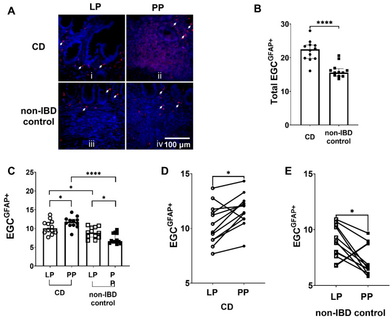
Enteric glial cells (EGC) expressing glial fibrillary acidic protein (EGCGFAP+) are more prominent in Crohn’s disease (CD). (A) Immunofluorescent staining of EGCGFAP+, i in lamina propria (LP) of CD patients, ii in Peyer’s patches (PP) of CD patients, iii in LP of non-inflammatory bowel disease (non-IBD) controls and iv in PP of non-IBD controls. The EGCGFAP+ are indicated by the arrows. Scale bar 100 μm. (B) Quantification of EGCGFAP+ in CD patients and non-IBD controls (C) The distribution of EGCGFAP+ in LP and PP of CD patients and non-IBD controls (D) The number of EGCGFAP+ was higher in PP of 11 out of the 12 CD patients analyzed, compared to LP (E) The number of EGCGFAP+ was lower in PP of 11 out of the 12 non-IBD controls analyzed, compared to LP. Data presented as median and interquartile range (IQR). Mann–Whitney U test was used for comparisons between groups and Wilcoxon matched-pairs signed-rank test for paired data, * p < 0.05, **** p < 0.0001.

