Figure 3.
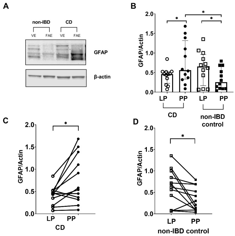
Glial fibrillary acidic protein (GFAP) levels are increased in tissue lysates of Crohn’s disease (CD) patients compared to non-inflammatory bowel disease (non-IBD) controls. (A) Representative Western blot image (converted into a black and white density blot) showing GFAP bands from one CD patient and one non-IBD control, respectively (B) Quantification of the bands after normalizing GFAP levels against the β-actin loading control (C) GFAP protein levels were higher in Peyer’s patches (PP) of 9 out of the 12 CD patients analyzed, compared to lamina propria (LP) (D) GFAP protein levels were lower in PP of 10 out of the 12 non-IBD controls analyzed, compared to LP. Data presented as median and IQR. Mann–Whitney U test was used for comparisons between groups and Wilcoxon matched-pairs signed-rank test for paired data, * p < 0.05.

