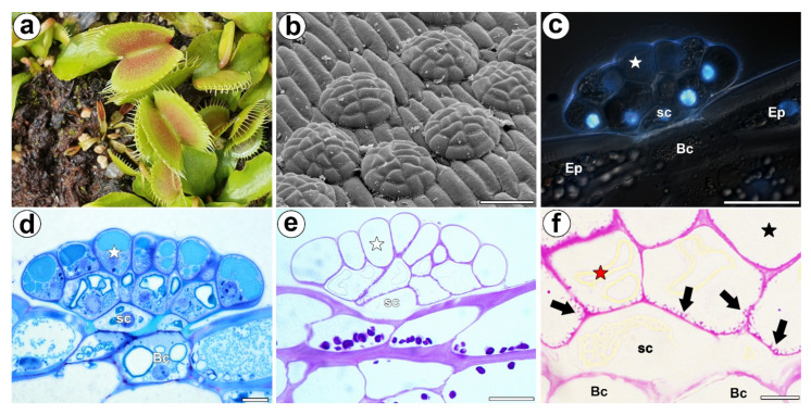
Figure 1.
Morphology of a D. muscipula trap. (a) Traps of D. muscipula. (b) Morphology of the digestive gland, bar 50 µm. (c) Structure of the digestive gland (stained with DAPI, blue fluorescence combined with Nomarski contrast): secretory cell (star), stem cell (sc), basal cell (Bc), epidermal cell (Ep), bar 20 µm. (d) A semi-thin section of the digestive gland: secretory cell (star), stem cell (sc), basal cell (Bc), bar 20 µm. (e) The digestive gland, PAS reaction: secretory cell (star), stem cell (sc), bar 20 µm. (f) The digestive gland, PAS reaction: cell wall ingrowths (black arrows), secretory cell from external layer of gland head (red star), secretory cell from inner layer of gland head (black star), stem cell (sc), basal cell (Bc), bar 2 µm.

