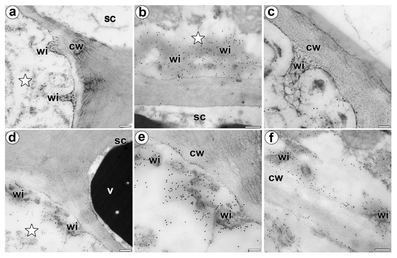
Figure 5.
Immunolocalization of the AGP in the cell walls of a D. muscipula digestive gland. (a) Immunogold labeling of the wall ingrowths with JIM14, a gland from an unfed trap: secretory cell (star), stem cell (sc), wall ingrowths (wi), cell wall (cw), bar 200 nm. (b) Immunogold labeling of the wall ingrowths with JIM14, a gland two hours after feeding: secretory cell (star), stem cell (sc), wall ingrowths (wi), bar 200 nm. (c) Immunogold labeling of the wall ingrowths with JIM8 in the secretory cell, a gland three days after feeding: wall ingrowths (wi), cell wall (cw), bar 100 nm. (d) Immunogold labeling of wall ingrowths with JIM13, in a gland three days after feeding: secretory cell (star), stem cell (sc), wall ingrowths (wi), cell wall (cw), vacuole (v), bar 200 nm. (e,f) Immunogold labeling of the wall ingrowths with JIM13 in the secretory cells, a gland three days after feeding: wall ingrowths (wi), cell wall (cw), bar 100 nm and bar 200 nm.

