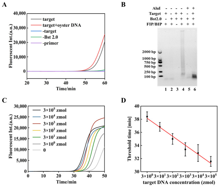
Figure 3.
Detection of V. parahaemolyticus by tRCA-lamp. (A) Real-time fluorescence analysis. [target DNA] = 3 amol (105 copies/μL), [FIP/BIP primers] = 1.6 μmol/L, [dNTP] = 1.4 mmol/L, 1 × SYBR Green II, 3.2 U Bst 2.0 DNA polymerase, 1 × isothermal amplification buffer at 65 °C for 60 min. (B) Endpoint product of tRCA-lamp with oyster DNA was digested with AluI and analyzed on 1% agarose gel electrophoresis. Lane 1–3, negative controls; lane 4, the product of tRCA-lamp; lane 5, 1 U AluI was added to the sample of lane 2; lane 6, 1 U AluI was added to the sample of lane 4. (C) Fluorescent intensity of tRCA-lamp under optimized conditions. [dNTPs] = 1.4 mmol/L, [primers] = 0.8 μmol/L, [MgSO4] = 6 mmol/L, 3.2 U Bst 2.0 DNA Polymerase, 1 × isothermal amplification buffer at 65 °C for 50 min. (D) Threshold time (Tt) was plotted against the lg[target concentration] of the reaction.

