Figure 2.
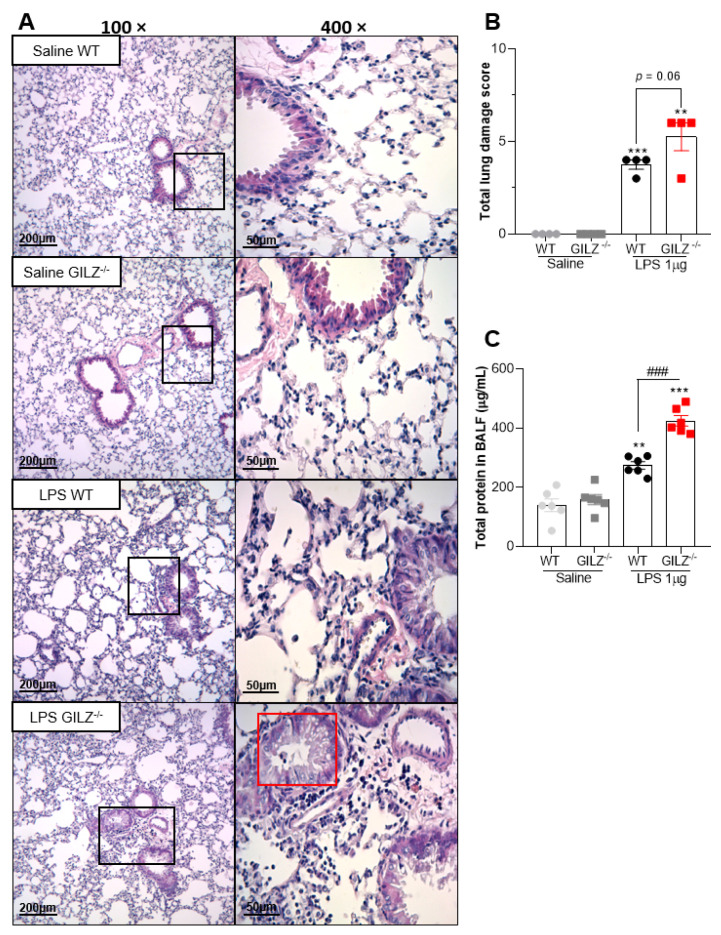
GILZ deficient mice show exacerbation of LPS-induced Acute Lung Injury. C57BL/6 WT or GILZ−/− mice were stimulated with LPS (1 µg, i.n.) and euthanized 24 h later. Representative slides of hematoxylin and eosin (H&E) stained lungs are shown (A). Scale bars = 200 µm (low magnification) and 50 µm (high magnification). Right slides are higher magnifications (400×) of the selected areas (boxes) in left slides (100×). Histopathological score evaluated airway, vascular, and parenchymal inflammation, neutrophilic infiltration, and epithelial injury (B). A red box represents bronchiolar epithelium degeneration, seen only in LPS-instilled GILZ−/− group of mice. The levels of total protein in BALF were evaluated (C). Data are mean ± SEM of N = 4 animals (histopathology) or N = 6 (protein levels) per group. ** p < 0.01 or *** p < 0.001 when compared to saline instilled groups; or as indicated: ### p < 0.001 by 2-way ANOVA or p = 0.06 when comparing LPS-challenged GILZ−/− to WT mice (t test).

