Figure 5.
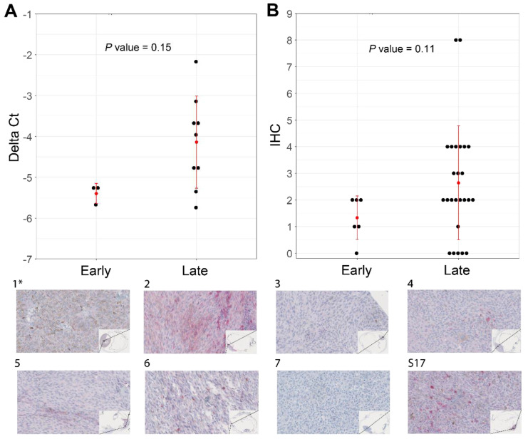
Increased levels of ABHD6 can be associated with SF3B1mut UM with late-onset metastatic disease (PFS ≥ 60 months). (A) RT-qPCR performed in triplicates using CHMP2A expression as a normalizer of 10 primary UM (see Supplementary Figure S5). (B) ABHD6 IHC on all SF3B1mut UM samples that were available and stratified for early- and late-onset metastatic disease. Red error bars represent standard deviation and red dot represents mean. Wilcoxon rank sum test was used to evaluate statistical difference of delta Ct values and IHC scores shown in scatterplots in panel A and B. p-value < 0.05 was considered statistically significant. (1*–7 and S17) ABHD6 IHC staining of eight primary UM samples, which are a selection of samples in panel A and B. The corresponding delta Ct values and IHC values with regard to histology are represented in Supplementary Figure S5. (40× magnification). (*) indicates the control sample with a PFS < 60 months.

