Figure 3.
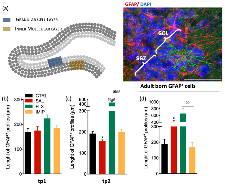
Morphological analysis of resident and newborn astrocytes in the hippocampal DG in a rat model of depression and after antidepressant treatment. (a) Representative scheme of the hippocampal DG regions where astrocytes were analyzed (left panel) and representative immunostaining and morphological analysis of GFAP+ cells in the hippocampal DG (right panel). (c,d) Longitudinal determination of the astrocytic length in the dorsal hippocampal dentate gyrus (dDG) in an experimental animal model of depression, at tp1 (b) and tp2 (c), specifically from GCL and IML. (d) Evaluation of astrocytic length in the granular cell layer (GCL) of the hippocampal DG newborn astrocytes, 4 weeks after cessation of the uCMS protocol and after treatment with fluoxetine and imipramine. These cells were identified by co-labeling GFAP+ and BrdU+ and were selected in the GCL to avoid stem cell analysis. * Represents uCMS effect analyzed by Student’s t-test. δ represents differences between ADs, analyzed by one-way analysis of variance (ANOVA). Data are represented as mean ± s.e.m. Scale bar represents 100 μm.* p < 0.05, δδ p < 0.01, ####, δδδδ p < 0.0001. Sample size: TP1: CTRL: 7; CMS: 10; FLX: 7; IMIP: 7; TP2: CTRL: 7; CMS: 10; FLX: 3–4; IMIP: 3–4; Adult-born GPAF+: CTRL: 5; CMS: 7; FLX: 5; and IMIP: 4. Abbreviations: GFAP, Glial Fibrillary Acidic Protein; CTRL, non-stressed animals; SAL, animals exposed to uCMS and injected with saline; IMIP, animals exposed to uCMS and treated with imipramine; FLX, animals exposed to uCMS and treated with fluoxetine; dDG, dorsal dentate gyrus, SGZ, subgranular zone; GCL, granule cell layer; tp1, time point 1 (6 weeks; immediately after the stress protocol cessation); and tp2, time point 2 (10 weeks; 4 weeks after the stress protocol cessation).

