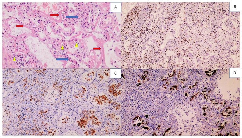
Figure 1.
Histology and immunohistochemistry of the lungs with diffuse alveolar damage (DAD). Photo (A) shows lymphocyte-infiltrated alveolar septa (blue arrows), hyaline membranes lining the alveolar septa (red arrows), and alveolar macrophages (yellow arrowheads), (HE, (hematoxylin-eosin staining) 200×). Photo (B) shows infiltration of immunohistochemically CD3-positive T lymphocytes in the alveoli (DAB (3, 3,-diaminobenzidine) positivity presented in brown, with blue contrast staining with hematoxylin 100×). Photo (C) shows numerous alveolar macrophages that are immunohistochemically CD68 positive (100×). Photo (D) shows numerous desquamated pneumocytes in the alveoli that are immunohistochemically AE1AE3 positive (100×).

