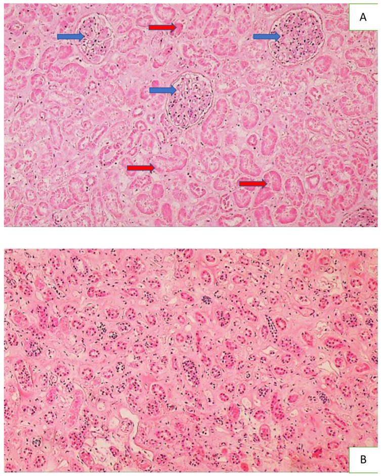
Figure 4.
Histology of the kidney of a COVID-19 patient with acute tubular necrosis ATN. Photo (A) shows acute tubular necrosis in the renal tissue, presented by the necrotic epithelium (no cells with nuclei present) of the proximal and distal convoluted tubules (red arrows) and the preserved glomeruli (blue arrows) (HE, 100×), while Photo (B) shows the preserved epithelium containing the nuclei (dark spots) of collecting tubules (HE, 100×).

