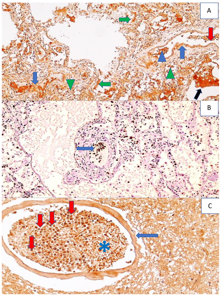
Figure 6.
Immunohistochemistry for 4-HNE protein adducts and MPO in the lungs and 4-HNE protein adducts in the brain of a COVID-19 patient. Photo (A) shows the immunohistochemically positive reaction to 4-HNE in the pulmonary blood vessel walls (blue arrows) and in the content of the blood vessel (red arrow), in the edematous fluid in the alveoli (black arrow) and hyaline membranes (green arrows), in some inflammatory cells in the blood vessels (blue arrowheads), and in alveolar macrophages (green arrowheads), (DAB, 100×). Photo (B) shows an immunohistochemically positive reaction to MPO, exclusively in neutrophils that are mainly in the blood vessels of the lungs (arrow) and rarely outside them in the lung tissue, presented as dark brown spots, (DAB, 200×). Photo (C) shows a strong immunopositive reaction to 4-HNE in a blood vessel wall (arrow), in the content of blood vessels (asterisk), especially in the neutrophils within the blood vessel (red arrows), while being sparsely present in the surrounding edematous brain tissue, visible as dark brown cells (DAB, 400×).

