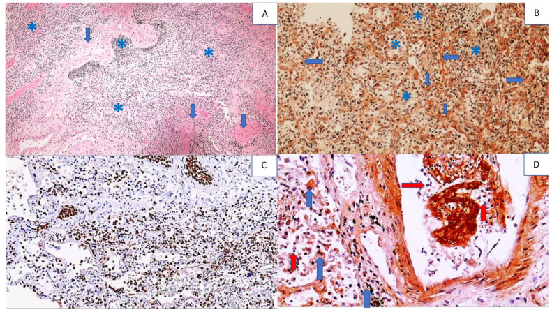
Figure 7.
Histology and immunohistochemistry of the lungs of a COVID-19 patient with pneumonia and DAD. Photo (A) shows the destruction of pulmonary tissue with a barely recognizable structure due to severe inflammation (asterisks) and fibrosis (arrows), (HE, 50×). Photo (B) shows the immunohistochemical positivity for 4-HNE in the alveolar septa (arrows), edematous fluid, alveolar macrophages, and other inflammatory cells (asterisks) (DAB, 100×). In contrast, in Photo (C), immunohistochemical positivity for MPO is seen only for inflammatory cells stained brown (DAB, 100×). Photo (D) shows strong immunohistochemical 4-HNE positivity in some inflammatory cells, e.g., alveolar macrophages (blue arrows), while other inflammatory cells, such as mononuclear cells, in the vessel and lung tissue are negative 4-HNE-negative (red arrows) (DAB, 400×).

