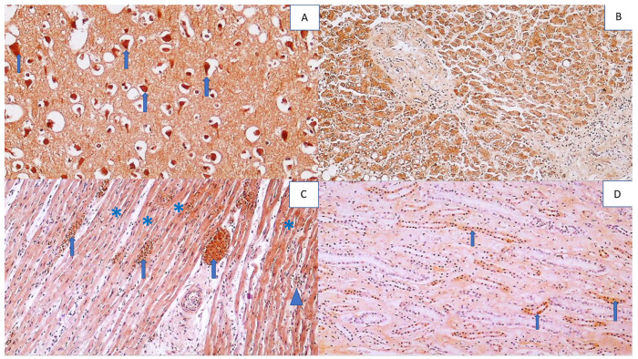
Figure 8.
Immunohistochemical positivity for the 4-HNE protein adducts in the vital organs (except the lungs, presented by other figures) of the deceased COVID-19 patient. Photo (A) shows very strong immunohistochemical positivity for 4-HNE in neurons (arrows) within the edematous brain tissue (DAB, 400×). Photo (B) shows a pronounced intensity (but weaker than that in neurons) of 4-HNE positivity in liver cells (DAB, 200×). Photo (C) shows moderate to strong 4-HNE positivity in myocardial muscle fibers affected by myocarditis (asterisks), with strong 4-HNE positivity in myocardial vasculature (arrows) and some inflammatory cells (arrowhead), (DAB, 200×). Photo (D) shows the weakest intensity of 4-HNE immunopositivity in the epithelium of the renal tubules (arrows) (DAB, 200×).

