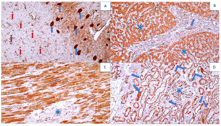
Figure 11.
Immunohistochemistry for SOD2 adducts in the vital organs of a COVID-19 patient (except the lungs). Photo (A) shows strong immunohistochemical positivity for SOD2 in neurons (blue arrows) and reactive astrocytes (red arrows) in brain tissue (DAB, 400×). Photo (B) shows strong immunohistochemical SOD2 positivity in hepatocytes (asterisks) and bile duct epithelium (arrow) (DAB, 200×). Photo (C) shows strong SOD2 positivity in myocardial muscle fibers, but not in the endomysium (asterisk) (DAB, 200×), while Photo (D) shows SOD2 positivity in renal tubules (arrows) and glomerulus (asterisk) (DAB, 200×).

