Figure 1.
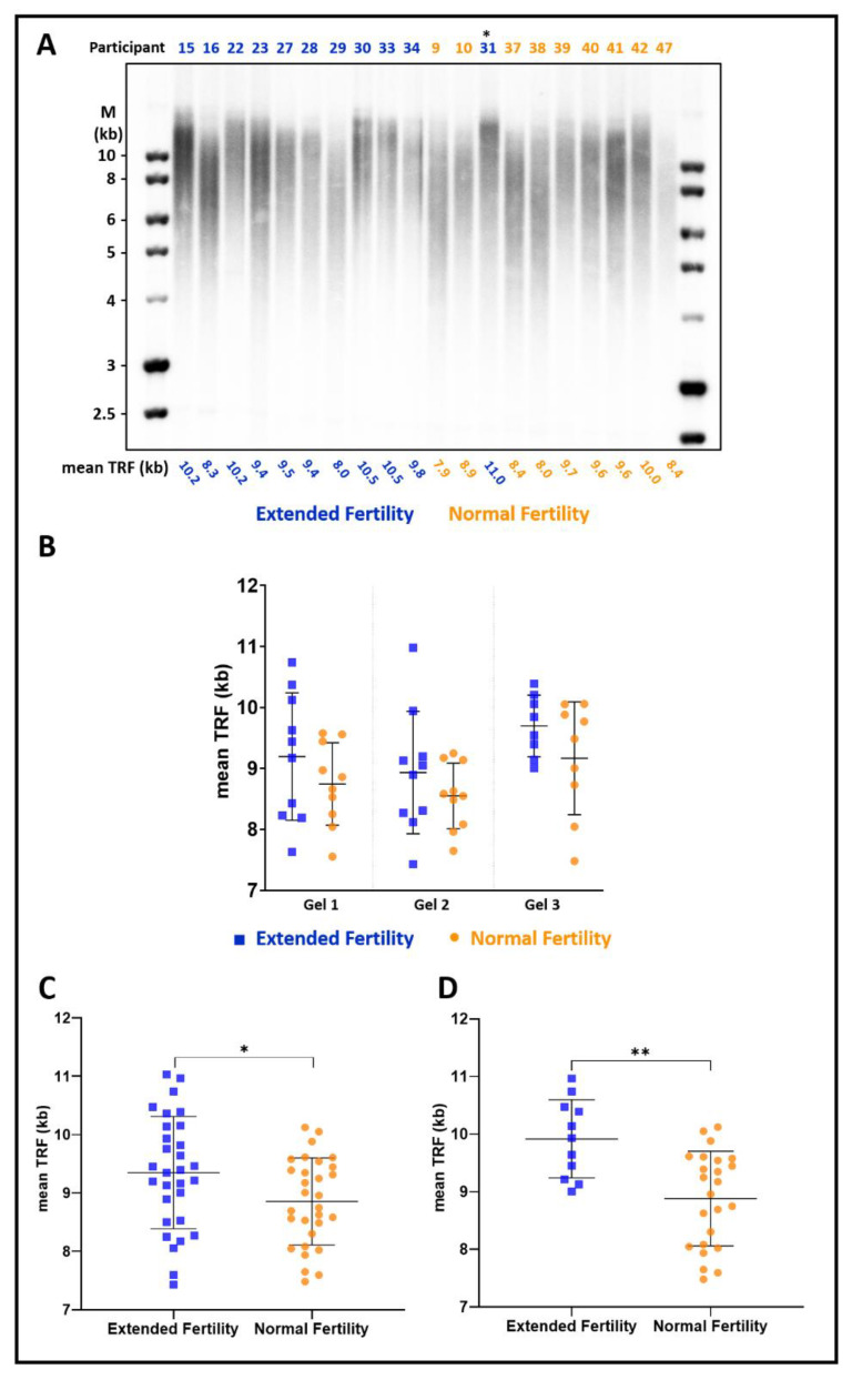
Women with extended fertility have longer telomeres. (A) A representative image of a Southern blot hybridized with a telomeric probe. Blood samples were collected from the Extended Fertility (EF) group participants (blue) within 48 h after delivery and the Normal Fertility (NF) group participants at recruitment (orange), and their leukocyte telomere length was analyzed as described under ‘Materials and Methods’. The mean telomere terminal restriction fragment (TRF) length, as calculated by TeloTool, is depicted below each lane. (B) Mean TRF in the NF and EF groups measured in three separate non-redundant gels shown in (A) and in the Supplementary Figure S1. Note that in (A), one of the women sampled as NF (indicated by an asterisk) later conceived and delivered a healthy child, thus reclassified as EF. (C) Mean TRF length reproducibly measured in several different gels for each participant, averaged, and presented for all the participants of the EF versus NF groups. p-value = 0.03 (*). Indicated are average and SD. (D) Mean telomere length is presented for a subgroup of participants with up to eight children in the EF and NF groups. p-value = 0.0009 (**). Indicated are average and SD. The graph for the subgroup of women with 9 or more children is shown in Supplementary Figure S3.

