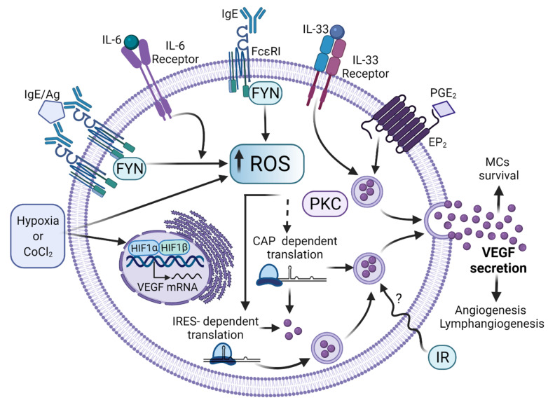
Figure 5.
Signaling pathways leading to VEGF synthesis and secretion in MCs. After distinct stimuli (some of them found in TME), MCs secrete VEGF to promote the formation of new blood vessels. Diverse ligands produced in TME or other conditions have been found to induce VEGF synthesis in MCs by controlling several steps on its synthesis and release. The binding of monomeric IgE to FcεRI and the antigen-dependent crosslinking of that receptor lead to ROS generation through the activation of Fyn tyrosine kinase, which promotes the accumulation of VEGF transcript and its translation through the internal ribosome-binding site (IRES) of VEGF mRNA. IL-33 and IL-6 receptors also lead to VEGF production in MCs, together with the triggering of the PGE2 receptor. Low-level ionizing radiation also leads to VEGF synthesis in MCs and this phenomenon leads to the restoring of blood vessels in damaged tissue. TME conditions, such as hypoxia or its mimicking agent cobalt chloride (CoCl2), lead to HIF-1α stabilization and promote VEGF transcription. Other intracellular pathways involved in VEGF synthesis in MCs require increased intracellular calcium levels and the activation of protein kinase C (PKC). Figure made in BioRender, agreement number FT23G75A9J.

