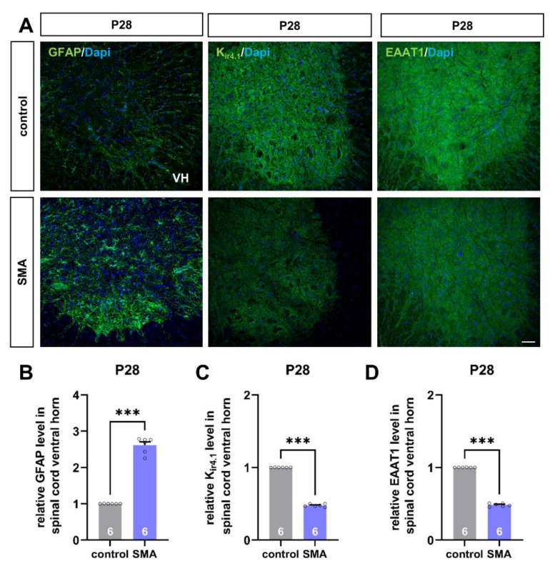
Figure 2.
Spinal astrocytes proteins are altered in a mouse model of late-onset SMA before the first loss of motor neurons. (A) Immunostaining of astrocyte-specific proteins such as GFAP (green), Kir4.1 (green), and EAAT1 (green) in spinal cord slices of SMA and control mice at P28. Nucleic DNA was stained with Dapi (blue). (B) The relative GFAP level in SMA mice spinal cord ventral horns was increased compared to control mice (***, p < 0.001). (C,D) The relative level of Kir4.1 and EAAT1 was reduced in SMA mice, compared to control animals (***, p < 0.001). VH = ventral horn. n = 6 animals (see bars). Three slices per spinal cord were investigated. Each data point reflects the mean of those three spinal cord slices. Scale bar: 20 µm.

