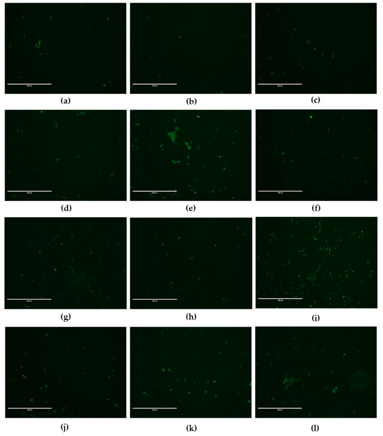
Figure 5.
Fluorescence microscopy images of JEG-Tox cells stained for caspase-3/7 activity. After 72 h incubation with (a) control, (b) DMSO, (c) ethanol, (d) bisphenol A at 20 µM, (e) diethylstilbestrol at 7.5 µM, (f) 4-tert-amylphenol at 50 µM, (g) 4-heptylphenol at 10 µM, (h) triclosan at 1 µM, (i) propylparaben at 100 µM, (j) benzyl butyl phthalate at 10 µM, (k) DEHP at 10 µM and (l) 3-benzylidene camphor at 10 µM, the cells were stained using Caspase-3/7 Green ReadyProbes™. Representative images from three independent experiments are shown. Bar scale: 400 µm. Magnification: 10×.

