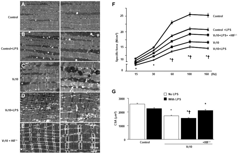
Figure 1.
Suppression of endotoxin-enhanced mechanical ventilation-induced diaphragm and mitochondrial injuries in HIF-1α-deficient mice. Representative micrographs of the longitudinal sections of diaphragm (×20,000: left panel; ×40,000: right panel) were from the same diaphragms of nonventilated control mice and mice ventilated at a tidal volume (VT) of 10 mL/kg (VT 10) for 8 h (n = 3 per group). (A,B) Nonventilated control wild-type mice with or without LPS treatment: normal sarcomeres with clear A bands, I bands, and Z bands; (C) 10 mL/kg wild-type mice without LPS treatment (normal saline): increase of diaphragmatic disarray; (D) 10 mL/kg wild-type mice with LPS treatment: disruption of sarcomeric structure, mitochondrial swelling with a vacuole-like structure, streaming of Z bands, and collection of lipid droplets; (E) 10 mL/kg HIF-1α deficient mice: reduction of diaphragmatic disruption. (F) Diaphragm muscle-specific force production was measured as described in Methods. (G) Cross-sectional area of diaphragm muscle fiber was measured as described in Methods (n = 5 per group). Mitochondrial swelling with concurrent formation of vacuoles and autophagosomes containing heterogeneous cargo are identified by arrows. * p < 0.05 versus the nonventilated control mice with LPS treatment; † p < 0.05 versus all other groups. Scale bar represents 500 nm. CSA = cross-sectional area; HIF−/− = hypoxia-inducible factor-1α-deficient mice; Hz = hertz; LPS = lipopolysaccharide; N = newton.

