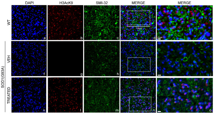
Figure 8.
Histone 3 acetylation in the lumbar spinal cord of male WT and SOD1(G93A) mice: The figure panel shows the different acetylation states of lysine 9 of histone 3 in the lumbar spinal cord of wild type (WT) mice, VEH and TREATED SOD1(G93A) groups. The nuclei were stained in blue with DAPI (a,f,k). The acetylation state of histone 3 was identified by the H3AcK9 antibody in red (b,g,l), while the motor neurons (MN) was detected by identifying the antibody neurofilament H with the SMI-32 antibody in green (c,h,m). The acetylation state of histone 3 was drastically reduced in the VEH group (g) compared to WT animals (b). The treatment with RESV and VPA led to a restoration of the acetylation of histone 3 in the TREATED group (l), (n = 3 WT, 5 VEH, 6 TREATED). Figure (d,i,n) shows the superimposed images of DAPI, SMI-32 and H3AcK9 in the WT, VEH and TREATED groups, respectively. Magnification of 20×, scale bar = 100 μm (a–d,f–i,k–n). The figure panel (e,j,o) shows the highlighted area in (d,i,n) at a higher magnification of 40×, scale bar = 10 μm.

