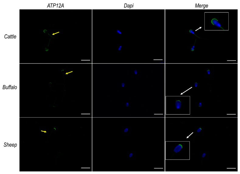
Figure 3.
ATP12A protein expression in sperm cells from cattle, buffalo and sheep. Representative fluorescence microscope images showing immunodetection of ATP12A (green) in frozen/thawed sperm cells. The secondary antibody was conjugated to Alexa Fluor 488. Nuclei were counterstained with DAPI (blue). Insets in the merge panels represent 2× magnification of representative sperm cells indicated by white arrows. Yellow arrows within the green channel indicate positive staining of ATP12A at the flagellum. Scale bar: 20 μm.

