Figure 1.
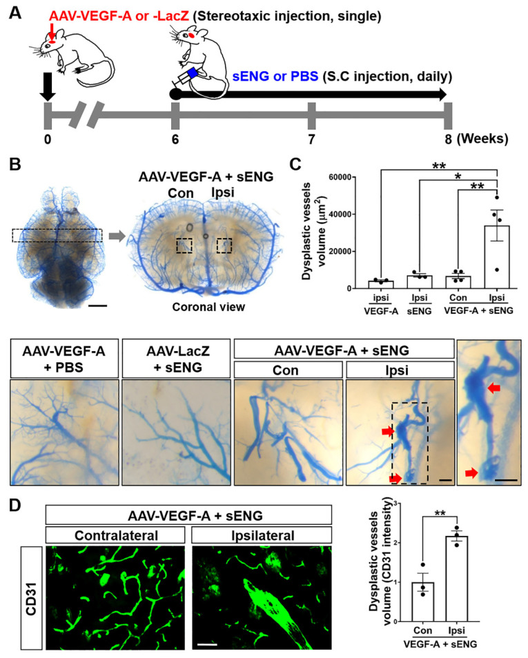
Soluble ENG/VEGF-A induces the formation of dysplastic vessels in the mouse brain. (A) Experimental scheme for sENG and AAV1-VEGF-A injection. The mice were stereotaxically injected with AAV1-VEGF-A (or AAV1-LacZ) into the intra-striatum and administered recombinant sENG (or PBS) subcutaneously (s.c.) every day for two weeks beginning at six weeks after the AAV1-VEGF-A injection. At eight weeks after the AAV1-VEGF-A injection, the mice were sacrificed. (B) Representative images of latex cast-clarified brains in mice injected with AAV1-VEGF-A + PBS, AAV1-LacZ + sENG, and AAV1-VEGF-A + sENG. Coronal sections showed the enlarged abnormal vasculature (inset, arrows) in the AAV1-VEGF-A injected site (ipsilateral) compared to the non-injected site (contralateral) in mice injected with AAV1-VEGF-A and sENG. Scale bars: 2 mm (whole brain image) and 100 μm (inset). (C) The quantification of vessel volume in mouse brains. AAV-VEGF-A (or VEGF-A), mice injected with vehicle (PBS) and AAV1-VEGF-A, sENG, mice injected with sENG and AAV1-LacZ, AAV-VEGF-A (or VEGF-A) + sENG, mice injected with AAV1-VEGF-A and sENG. n = 3–4, * p < 0.05, ** p < 0.01, One-way ANOVA. (D) Image of CD31 immunostained brain from mice injected with sENG and AAV1-VEGF-A and the quantification of CD31 intensity. Scale bar: 50 µm, n = 3, data are presented as mean ± SEM, ** p < 0.01, Student’s t-test. Con, contralateral, Ipsi, ipsilateral.

