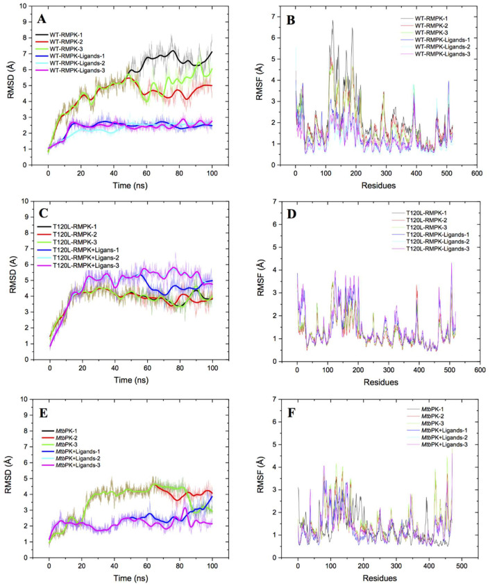
Figure 7.
RMSD and RMSF from molecular trajectories of WT-RMPK (A,B), T120L mutant (C,D), and MtbPK (E,F). Initial structures were in the presence of Mg2+, PEP, Mg-ADP and K+ for the K+-dependent WT-RMPK and T120L mutant and without K+ for the K+-independent MtbPK. The presence or absence of ligands are indicated by different colors in (A,C,E), whereas red and blue lines indicated with and without ligands in (B,D,F). Triplicates of molecular dynamics were carried out and the analyzes were made with CPPTRAJ [32]. For the particular case of MtbPK, runs 1 and 2 overlapped, either with or without ligands (E).

