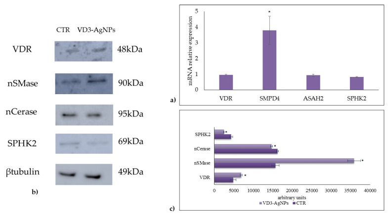
Figure 4.
Effect of VD3 + AgNPs on gene and protein expression in HaCaT cells. Cells were cultured and treated as reported in Section 4. (a) Gene expression evaluated by RT-PCR; glyceraldehyde 3-phosphate dehydrogenase and 18S rRNA were used as housekeeping genes. (b) Western blotting; the position of proteins was indicated in relation to the position of molecular size standards, β-tubulin was used as loading control. (c) Area density evaluated by Chemidoc Imagequant TL software. VDR, vitamin D3 receptor; nSMase neutral sphingomyelinase; nCerase, neutral ceraminidase; and SPHK2, sphingosine kinase 2. Values of proteins, normalized with β-tubulin, are expressed as arbitrary units in control and experimental sample (treated with 133 nM VD3 + 5 ppm AgNPs) and represent the mean ± SD of three independent experiments performed in duplicate. Significance, * p < 0.05 versus the control sample.

