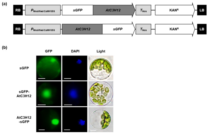
Figure 3.
Subcellular localization of AtC3H12. (a) Schematic maps of the sGFP-fused, full-length ORF of AtC3H12 constructs. (b) Subcellular localization of AtC3H12 protein investigated by transient expression of sGFP-AtC3H12 and AtC3H12-sGFP constructs in Arabidopsis protoplasts. Left, GFP signal; middle, 4′,6-diamidino-2-phenylindole (DAPI) staining; right, light microscopic picture. The white lines indicate scale bar = 10 μm.

