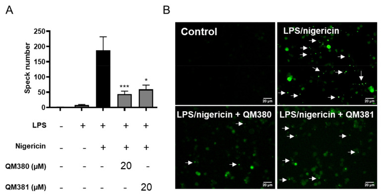
Figure 4.
QM380 and QM381 inhibit ASC speck formation: (A) Percentage of ASC specks measured in THP-1-ASC-GFP cells treated with QM380 and QM381 (20 µM) and stimulated with LPS (100 ng/mL) and nigericin (10 µM). (B) Live-cell imaging of THP-1-ASC-GFP cells treated as indicated above. Scale bar corresponds to 20 µm. Arrows point to ASC specks. Asterisks represent significant differences compared to the stimulated control (LPS/nigericin) as determined by a one-way ANOVA test with Tukey’s multiple post-test comparisons * p < 0.05; *** p < 0.001. All data are expressed as the mean ± SD of four independent experiments.

