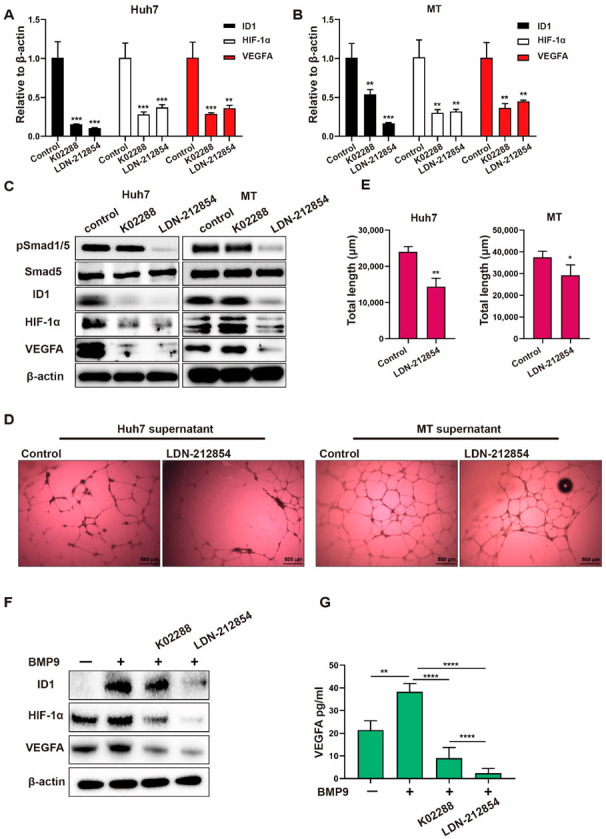
Figure 6.

BMP receptor inhibitors repress BMP9-induced HIF-1α/VEGFA signaling activity in HCC cells. (A,B) Relative gene expression levels of ID1, HIF-1α and VEGFA in Huh7 and MT cells. Cells were harvested after being treated with 1 μM BMP receptor inhibitors for 48 h. (C) Western blot analysis of pSmad1/5, Smad5, ID1, HIF-1α and VEGFA in Huh7 and MT cells. Cells were harvested after being treated with 1 μM BMP receptor inhibitors for 48 h. (D,E) Lumen formation assay of HUVECs treated with supernatants from Huh7 and MT cells for 24 h. Cell supernatants were harvested following treatment with 1 μM BMP receptor inhibitors or 10 nM ramucirumab. (F) Western blot analysis of ID1, HIF-1α and VEGFA in Huh7 cells treated with 1 μM BMP receptor inhibitors in the presence of BMP9 (5 ng/mL) for 48 h. (G) ELISA analysis of VEGFA in Huh7 supernatants. Supernatants were collected after cells were treated with 1 μM BMP receptor inhibitors in the presence of BMP9 (5 ng/mL) for 48 h. The error bars represent the SD from at least three independent biological replicates. Student’s t test was used to calculate p values, represented as * p < 0.05; ** p < 0.01; *** p < 0.001; **** p < 0.0001.
