Figure 1.
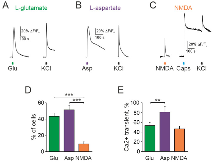
Intracellular Ca2+ transients activated by glutamate receptors agonists in rat TG neurons: Representative Ca2+ traces recorded after (A) glutamate (1 mM), (B) aspartate (100 µM), and (C) NMDA (100 µM) applications to TG neurons. All agonists were mixed with the co-agonist glycine (10 µM) in magnesium free solution. Average of 5 traces in each. Notice that NMDA sensitive neuron also responded to the TRPV1 agonist capsaicin; (D) Histograms showing the percentage of neurons responding to three glutamate agonists. Notice that the number of neurons responding to NMDA was significantly less than to glutamate and aspartate; (E) Histograms showing amplitudes of Ca2+ transients (normalized to ionomycin response) activated by glutamate, aspartate, NMDA. All agonists were applied with the co-agonist glycine. Mean ± SEM. ** p < 0.01; *** p < 0.001.

