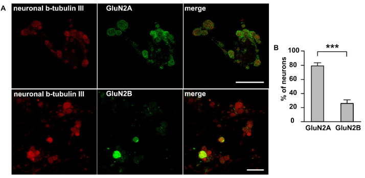
Figure 3.
Immunolabeling NMDA receptors in trigeminal ganglion neurons: (A) Immunostaining of GluN2A and GluN2B subunits of NMDA receptor in TG cells; Left column—labelling of β-tubulin III; central column—labelling with GluN2A (top) or GluN2B (bottom) antibodies; right column—overlay. Representative images of the staining are made at original magnification 63 × 1/4. Scale bar: 50 μM (B) Histogram presented the percentage of neurons expressed GluN2A and GluN2B subunits, *** p < 0.001.

