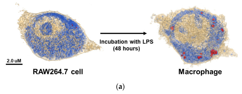
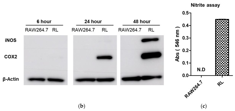
Figure 1.
(a) The 3D holotomographic images show RAW264.7 cells and LPS-induced macrophages by RI-based imaging technology. When RAW264.7 cells differentiated into macrophages, lipid droplets appeared inside the cells. (Red dots indicate lipid droplets) (b) Time-dependent protein expression level of LPS-induced macrophages were analyzed by Western blotting. iNOS and COX2 proteins showed that LPS was activated into RAW264.7 cells to induce macrophages. Β-actin was used as a control. (c) Nitrite production in the cell media under LPS treatment was measured by the Griess assay. Nitrite production indicates that iNOS protein induces inflammation in macrophages. (RL: LPS treated RAW264.7 cell).


