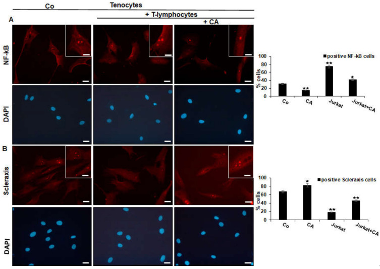
Figure 3.
Effect of Calebin A (CA) on NF-κB (A) and on Scleraxis (B) in tenocytes cultured in an inflammatory environment analyzed by immunofluorescence. Serum-starved tenocyte monolayer cultures alone (control, Co) or co-cultured with T-lymphocytes were either left untreated or treated with CA (5 µM) for 5 h. Magnification ×400; scale bar = 45 nm. All experiments were conducted three times and quantification of positively NF-κB- or Scleraxis-labeled nuclei was performed by counting 400–500 cells from 10 different microscopic fields. Values of p < 0.05 (*), p < 0.01 (**) were considered statistically significant in relation to control.

