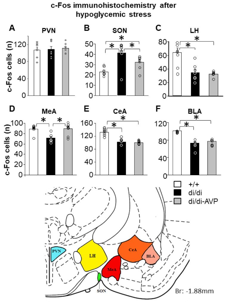
Figure 4.
The number of c-Fos immunoreactive cells (individual values = circles and means + SEM) counted in defined brain areas 60 min after insulin injection in male Brattleboro rats after AVP synthesis rescue. Unlike in the PVN (A) di/di animals showed a higher number of c-Fos positive cells in the SON (B) and a reduced number in LH, (C) MeA (D), CeA (E) and BLA (F) than +/+. Please note that in di/di-AVP animals, this difference was normalized in the SON and MeA only. The schematic drawing obtained from a rat brain atlas illustrates the reference brain areas selected for the microscopic analysis (n = 7–8/group). Abbreviations: AVP, vasopressin; BLA, basolateral amygdala; CeA, central amygdala; di/di, vasopressin-deficient Brattleboro rat; di/di-AVP, di/di animals with vasopressin synthesis rescue in the supraoptic nucleus; LH, lateral hypothalamus; MeA, medial amygdala; PVN, paraventricular nucleus of the hypothalamus. * p < 0.05, one-way ANOVA followed by Newman–Keuls comparisons.

