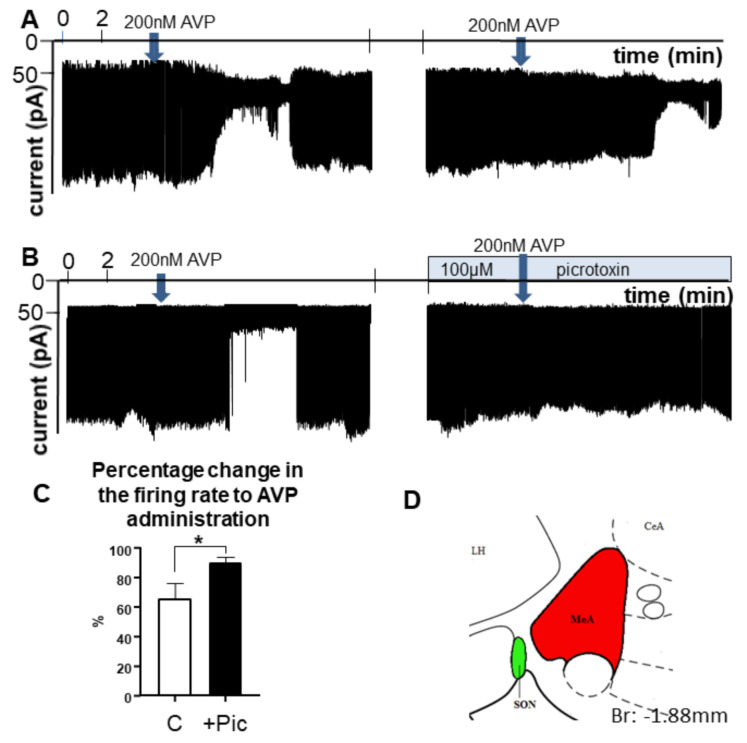
Figure 5.
Representative examples of electrophysiological recordings illustrating the effect of AVP on the firing of MeA neurons in a brain slice obtained from +/+ rats. (A) Approx. 1–2 min after administration of 200 nM synthetic, AVP firing of MeA neurons was reduced. Repeated application of AVP resulted in a similar decrease. (B) The effect of AVP could be eliminated by the GABAA-R blocker picrotoxin. (C) The bar graph summarizes the results. The arrow indicates the time of application of the single bolus of AVP. (D) The inset shows the area of the slice that was chosen for the recording (n = 10). * = p < 0.05, Student’s t-test. Abbreviations: AVP, vasopressin, C: control, only AVP was administrated; MeA, medial amygdala; Pic, picrotoxin.

