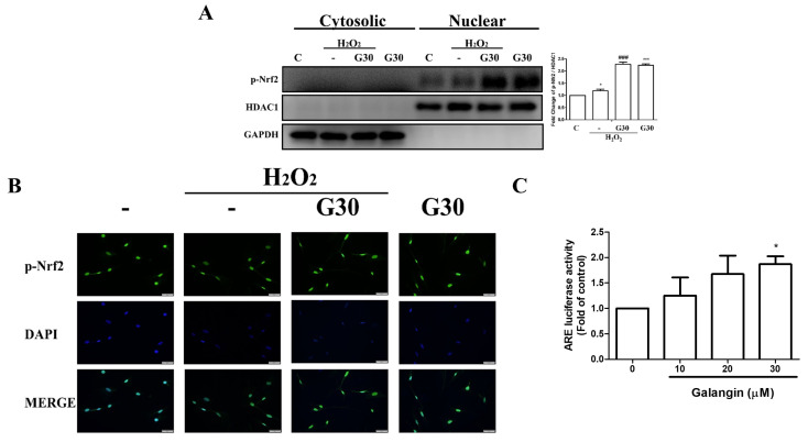
Figure 3.
Galangin triggers Nrf2 nuclear translocation and ARE transcriptional activation in HS68 cells exposed to H2O2. (A,B) HS68 cells were exposed to H2O2 (200 μM) or UVB (40 mJ/cm2) and then co-treated with galangin at a specific concentration (30 μM). p-Nrf2 expression was estimated in the cytosolic and nuclear fractions. The p-Nrf2 protein level was detected by Western blot. (C) p-Nrf2 nuclear translocation was further ascertained by immunofluorescence staining. An anti-Nrf2 antibody and a FITC-conjugated second antibody were used to detect p-Nrf2 cellular distribution. DAPI staining indicated the nucleus location (blue). The images were acquired using florescence microscopy (200×). HS68 cells were transfected with ARE-luciferase construct for 24 h. The cells were then treated with different doses of galangin (10, 20, and 30 µM) for 24 h and assayed for luciferase activity. Values shown are means ± SE. Quantification of the results is shown (n = 3) * p < 0.05, *** p < 0.001 vs. untreated control cells; ### p < 0.001 vs. H2O2-treated cells.

