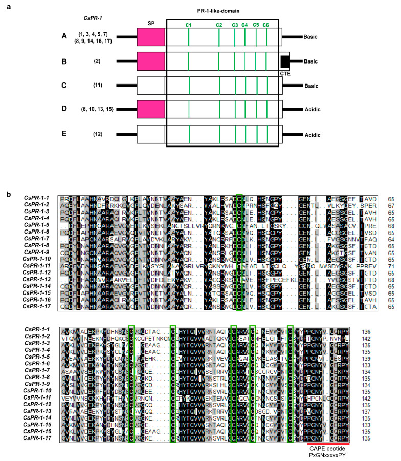
Figure 1.
Genomic and domain structures of the CsPR-1 genes. (a) Diagram of the genomic structures of the CsPR-1 genes. Open boxes signify the open reading frames (ORFs). Signal-peptide (SP) regions are shaded in pinkish red. Vertical green solid bars represent the positions of the six conserved cysteine residues (C1-C6). The box drawn with black solid lines indicates the conserved PR-1-like domains (pfam cd05381). The interior black box indicates the C-terminal extension (CTE). (b) Amino-acid alignment of the PR-1-like domain of the deduced CsPR-1 proteins. DNAMAN 7.0 was used to mark the amino-acid residues with an identity of more than 50% with black shade. The areas in the interior green boxes and the red solid line indicate the C1-C6 and CAPE peptide, respectively.

