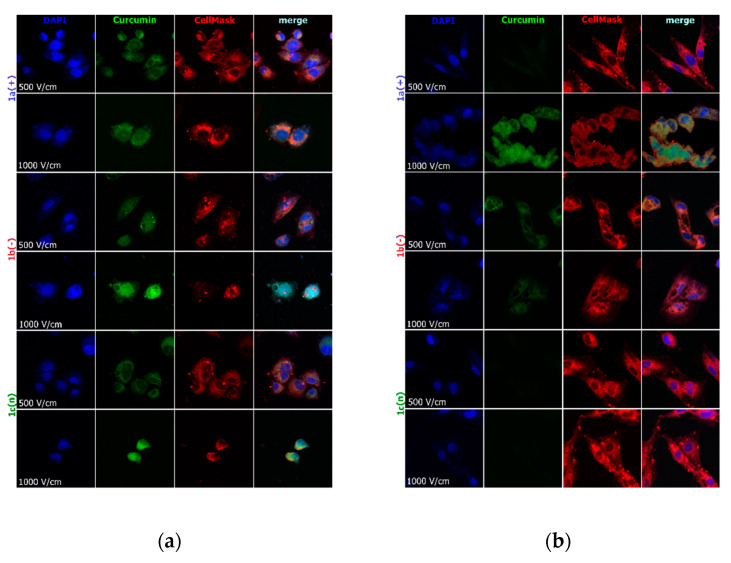
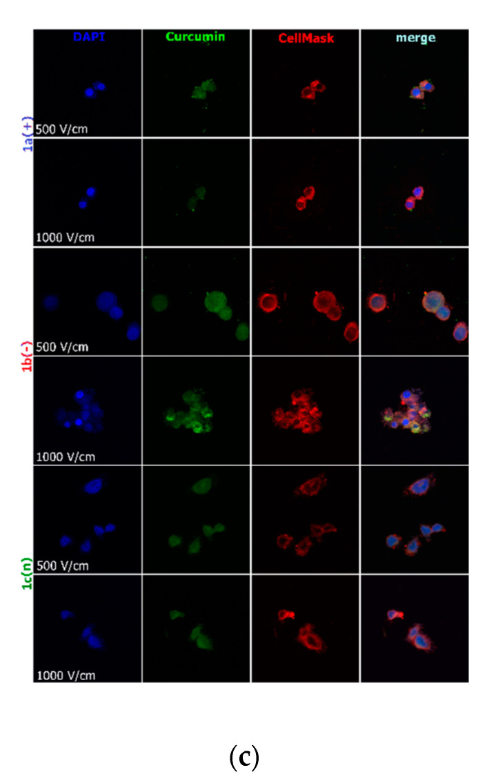
Figure 4.
Cells electroporated (500 and 1000 V/cm) with encapsulated curcumin and stained for nuclei (DAPI) and cell membrane (CellMask DeepRed) visualization: (a) hamster ovarian fibroblasts (CHO-K1 cells); (b) normal rat skeletal muscle cells (L6 cells); (c) colon adenocarcinoma cells (LoVo cells). CUR concentration was 4 μM.


