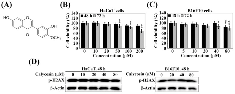
Figure 1.
Cytotoxicity of calycosin in HaCaT and B16F10 cells. (A) Chemical structure of calycosin. (B) Human keratinocyte HaCaT cells and (C) mouse melanoma B16F10 cells were treated with indicated concentrations of calycosin for 48 and 72 h. Cell viability was analyzed by MTT assay. Data are represented as the means ± S.D. from three independent experiments. Significant difference versus control: * p < 0.05, ** p < 0.01. (D) Western blotting analysis of p-H2AX expression in HaCaT (left) and B16F10 (right) cells treated with indicated concentrations of calycosin for 48 h.

