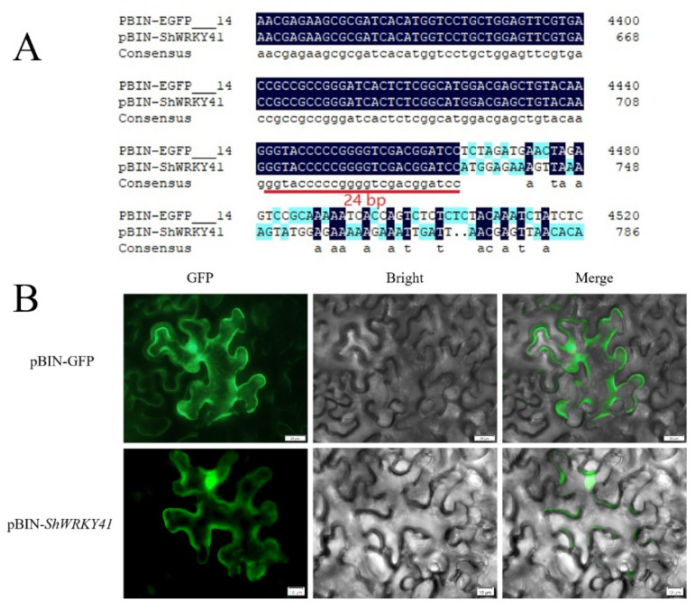
Figure 6.
ShWRKY41 located in plasma membrane and nucleus. (A) The sequence analysis of pBIN-ShWRKY41 vector by Sanger sequencing. The 24 bp gap means that the GFP coding sequence and CDS of ShWRKY41 did not exist frameshift mutation. (B) Images were collected by confocal microscopy at 24 h post pBIN-ShWRKY41 inoculation. Green fluorescent protein (GFP) signal was visualized by confocal microscopy.

