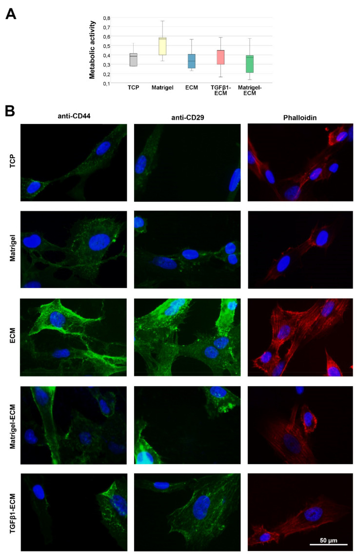
Figure 2.
Metabolic activity and microscopy. (A) Metabolic activity of MSC after culture on the different MSC-derived ECM substrates (ECM, Matrigel-ECM, TGFβ1-ECM) or in the respective control conditions without MSC-derived ECM (tissue culture plastic (TCP), Matrigel), as determined by MTS-assay (n = 5 independent experiments). (B) Representative images of surface molecule (CD44, CD29) and actin cytoskeleton (phalloidin) staining of MSC cultured on the different substrates. Note the higher intensity of the stainings in MSC cultured on ECM, indicating that the surface molecules interacting with the ECM are more present and the cytoskeleton formation is more pronounced.

