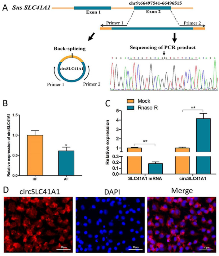
Figure 1.

Identification and validation of circSLC41A1. (A) Sketch of the structure of circSLC41A1, which is generated from the SLC41A1 gene exon 2 via back splicing. (B) SLC41A1 mRNA and circSLC41A1 expression with and without RNase R digestion. Untreated GAPDH was used as an internal control. (C) Differential expression of circSLC41A1 in HFs and AFs detected by qRT-PCR (n = 8). (D) The cytoplasmic localization of SLC41A1 in GCs detected by FISH. SLC41A1 was labeled with red fluorescence, and the nuclei were stained by DAPI (blue). Scale bar: 20 μm. Data are expressed as the mean ± SEM, * p < 0.05, ** p < 0.01.
