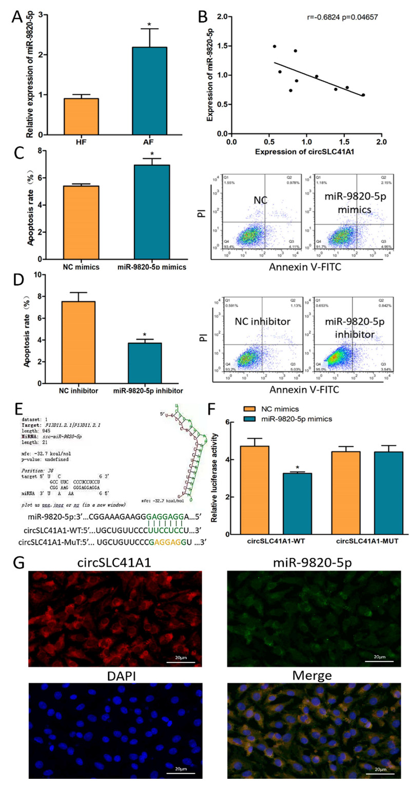
Figure 3.

circSLC41A1 is a possible sponge for miR-9820-5p. (A) Differentially expressed miR-9820-5p in HFs and AFs detected by qRT-PCR. (B) Negative correlation between circSLC41A1 and miR-9820-5p expression levels in individual follicles (n = 9). (C,D) Shift of GC apoptosis rates after miR-9820-5p mimics and inhibitor transfection detected by flow cytometry. (E,F) The binding of miR-9820-5p of circSLC41A1 was verified by dual-luciferase activity analysis. (G) The subcellular co-localization of circSLC41A1 (labeled by red fluorescence) and miR-9820-5p (labeled by green fluorescence) in GCs verified by FISH. The nuclei were stained by DAPI (blue). Scale bar: 20 μm. Data are expressed as the mean ± SEM of three experiments; * p < 0.05.
