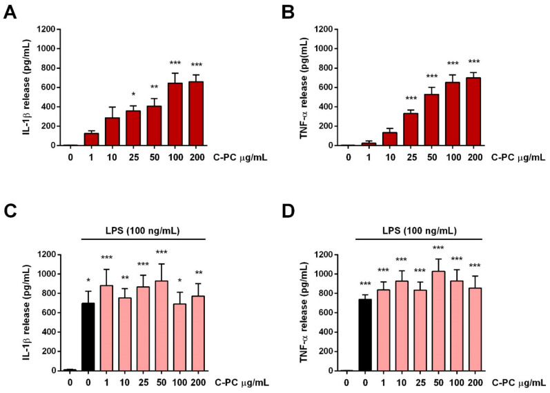
Figure 2.
Effect of C-phycocyanin on cytokine release from cortical microglia. Microglia were cultured overnight medium containing 10% of serum, which was replaced with serum-free medium before treatment with C-PC (A,B) or C-PC + LPS (100 ng/mL) (C,D). After the exposure to C-PC or LPS for 16 h, supernatants were collected and analyzed for IL-1β (A,C) and TNF-α (B,D) content. Data are means ± SEM (n = 3 in triplicate). * p ˂ 0.05, ** p ˂ 0.01, and *** p ˂ 0.001 versus control cells. One-way ANOVA followed by Holm–Sidak’s test.

