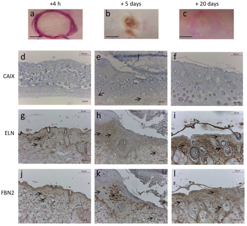
Figure 6.
Effect of pressure-induced hypoxic ulcer model on ELN and FBN2 fibers in mice. Representative images of mouse dorsal skin at (a) 4 h, (b) 5 days and (c) 20 days after removal of magnet compression. Corresponding skin immunohistochemistry for (d–f) CAIX, (g–i) ELN and (j–l) FBN2. Scale bars = (a–c) 4 mm, (d–f) 100 µm, and (g–l) 50 µm.

