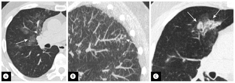
Figure 6. Pulmonary infection. In A, axial image of the right middle lobe, showing focal areas of ground-glass attenuation (arrow) with ill-defined nodularity. In B, a tree-in-bud pattern (arrow). The patient had clinically and microbiologically confirmed pneumonia. In C, axial image of a different patient, showing consolidation with air bronchogram in a predominantly peribronchovascular distribution (arrow). The patient was clinically diagnosed with bronchopneumonia.

