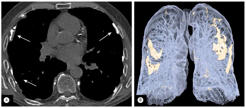
Figure 7. Pleural plaques. In A, axial image with bone window settings, showing several linear calcifications in the costal pleura (arrows). In B, 3D reconstruction of the same patient, showing that the pleural plaques are also affecting the mediastinal pleura and the diaphragmatic pleura.

